
Crazy 1991 MRI Scan Study Shows What Happens to the Body During Intercourse
Intercourse has always been surrounded by curiosity, myths, and assumptions about how the human body behaves during intimacy. For decades, scientists had theories about what happens internally, but they lacked direct visual evidence. That changed when an unusual scientific experiment placed a couple inside an MRI scanner while they engaged in intercourse. The goal was simple but ambitious: capture real images of the human body during intercourse and understand what actually happens inside.
The experiment, which first took place in the early 1990s, produced some of the most fascinating medical images ever recorded. It revealed unexpected details about anatomy, challenged old beliefs that had existed for centuries, and sparked global interest in how intercourse really works inside the human body.
The story behind this research began with Dutch scientist Menko Victor “Pek” van Andel. He wanted to investigate the mechanics of intercourse using modern imaging technology. At the time, MRI scanners were still relatively new tools, and researchers were just beginning to explore their potential for studying different parts of the body.
Van Andel decided that an MRI scan could capture something that had never been seen before. If a couple were willing to participate, the scanner could record real images of intercourse and show exactly how male and female anatomy align during the act.
The volunteers were Ida Sabelis and her partner Jupp, who agreed to take part in the unusual experiment. They entered the MRI machine while researchers observed and captured images from the control room. Their willingness to participate helped scientists obtain the first visual documentation of intercourse inside the body.
Although intercourse is a natural part of human life, surprisingly little direct research has examined the physical mechanics involved.
Much of what scientists believed about anatomy during intercourse was based on speculation, drawings, and older medical theories.
For centuries, influential figures such as Leonardo da Vinci believed that the male anatomy remained straight during intercourse. These ideas shaped the way doctors and researchers thought about sexual anatomy. However, without modern imaging technology, there was no clear way to verify whether those assumptions were correct.
The MRI experiment allowed scientists to observe intercourse in a completely new way. Instead of relying on diagrams or theory, they could see real images showing how the body behaves during intercourse.
Carrying out the study was not easy. MRI machines are narrow, loud, and designed for medical imaging rather than comfort. Participants usually have to lie still while the scanner captures images.
For the couple involved in the study, maintaining position during intercourse inside the machine was challenging. The scanner produced loud noises and provided very little space to move.
Researchers also had to give instructions from the control room while the scans were taken. Because early MRI technology captured images slowly, the couple sometimes had to remain still for several seconds so the machine could record the necessary data.
Despite these obstacles, the experiment succeeded in capturing detailed images of intercourse inside the human body.
Another limitation came from the physical size of the MRI scanner. The narrow tube meant the couple could not move freely or choose different positions.
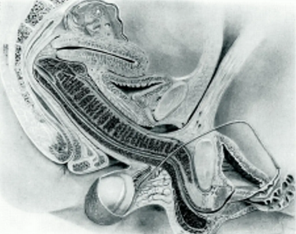
The researchers initially hoped to capture images of intercourse in the missionary position. However, the restricted space inside the machine made that impossible.
Instead, the couple used a spooning position so they could fit inside the scanner while still allowing the machine to capture clear images. Even this arrangement required careful positioning to ensure the scan would work properly.
Although the setup was far from romantic, it allowed scientists to document intercourse in a way that had never been done before.
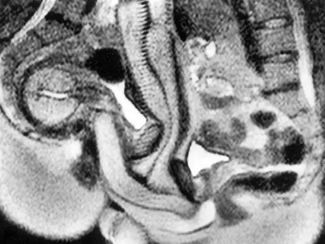
The images produced during the experiment provided remarkable insight into how the body works during intercourse. One of the most surprising discoveries involved the shape of the male anatomy during penetration.
Instead of remaining straight, the images showed that the penis bends inside the vaginal canal. The shape closely resembled a curved or boomerang form. This bending allows the anatomy of both partners to align naturally during intercourse.
This finding contradicted centuries of assumptions that had appeared in early medical drawings and anatomical studies. It also demonstrated that the body adjusts in complex ways during intercourse to accommodate natural movement and positioning.
The MRI scans also revealed new information about how the female body responds during intercourse. Researchers observed changes in the uterus and surrounding structures when arousal occurred.
One key observation showed that during arousal without intercourse, the uterus rises and the front wall of the vagina lengthens. These adjustments prepare the body for intercourse and help accommodate penetration.
However, the scans indicated that the uterus itself does not increase in size during intercourse. This detail helped clarify how female anatomy behaves during the process and corrected several misunderstandings that had circulated in earlier medical discussions.
The initial experiment produced valuable information, but researchers wanted to gather more data. Over the following years, the study expanded to include additional volunteers.
Between 1991 and 1999, eight couples and three individual women participated in similar MRI experiments. In total, the machine captured images of intercourse thirteen more times.
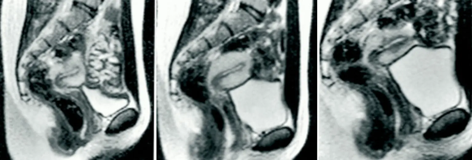
Unlike the original couple, most participants attempted intercourse in the missionary position during the scans. Because the environment inside the MRI machine was not particularly comfortable or romantic, male participants were required to take medication to help maintain arousal during the study.
While analyzing the scans from these experiments, researchers noticed something unusual. In every recorded case, the woman’s bladder appeared to fill rapidly during intercourse.
This happened even when participants had used the bathroom shortly before entering the MRI machine. By the end of the scans, the images consistently showed a noticeably full bladder.
Scientists were intrigued by this observation because they did not fully understand why it occurred. The pattern appeared repeatedly across multiple participants and different scans.
Although the exact reason remains unclear, researchers proposed a possible explanation for the bladder behavior observed during intercourse.
Van Andel suggested that the body might be responding in a way that encourages urination after intercourse. One theory is that this mechanism could help reduce the risk of urinary tract infections by flushing bacteria from the urinary tract.
However, this idea remains theoretical. Scientists have not confirmed the exact cause of the bladder filling observed during intercourse in the MRI scans.
For Ida Sabelis, participating in the experiment was a unique experience that she later described in interviews. She explained that the process was not particularly romantic, largely because of the clinical environment and the technical instructions from researchers.
The couple had to remain in position while the scanner captured images, which sometimes meant staying still for extended moments.
Despite the awkward conditions, Sabelis later reflected positively on the experiment. She recognized that their participation helped scientists learn more about the mechanics of intercourse and contributed to medical understanding.
When the research findings were eventually published in the British Medical Journal in 1999, the images quickly attracted attention.
They became some of the most widely discussed visual examples of human anatomy during intercourse. The images were unusual not only because of their subject matter but also because they used advanced medical imaging to show real internal alignment between male and female anatomy.
Over time, the study became widely known among medical professionals, researchers, and curious readers who wanted to understand how intercourse works from an anatomical perspective.
@viceau Sh@gging in an MRI machine for science #australia #themoreyouknow #science #peach #eggplant #seggs ♬ original sound – VICE Australia
The unusual nature of the experiment naturally sparked strong reactions. Some people were surprised that scientists had conducted such a study at all. Others were fascinated by the insight it provided into the human body.
Initially, the research faced criticism and skepticism. Some individuals questioned whether the experiment was necessary or appropriate.
However, as interest in the findings grew, attitudes began to shift. The images and data generated by the study became valuable references for understanding anatomy during intercourse.
Before the MRI experiment, much of what scientists believed about intercourse was based on outdated assumptions. The new images provided clear visual evidence that corrected several misconceptions.
One of the most important discoveries involved the curved shape of the penis during intercourse, which contradicted earlier beliefs that it remained straight.
The scans also helped researchers understand how the female body prepares for intercourse through internal adjustments that make penetration possible and comfortable.
Even decades after the original experiment, the study continues to attract attention. Researchers and educators still reference the images when discussing the mechanics of intercourse and human anatomy.
The project demonstrated how modern medical imaging can reveal details about the body that were previously impossible to observe. It also highlighted how scientific curiosity can lead to discoveries in unexpected areas of research.
Although the experiment may sound unusual, it provided valuable insight into a fundamental aspect of human biology.
More than thirty years after the first scans were captured, the MRI images of intercourse remain widely discussed. They continue to appear in articles, documentaries, and educational material exploring human anatomy.
Part of the reason the study remains popular is the unusual method used to capture the data. Few scientific experiments have involved participants engaging in intercourse inside medical imaging equipment.
At the same time, the research answered questions that had puzzled scientists for generations. By visualizing intercourse directly, the MRI scans offered a clearer understanding of how the body functions during intimacy.
Intercourse was once an area of anatomy surrounded by speculation and incomplete knowledge. The MRI experiment involving Ida Sabelis and her partner changed that by capturing real images of the body during intercourse.
Through this unusual study, researchers discovered new details about how male and female anatomy align, how the body responds internally, and how certain physical processes occur during intercourse.
Although the experiment took place decades ago, its findings still influence discussions about human anatomy today. What began as a bold scientific idea ultimately provided a clearer picture of intercourse and the remarkable ways the human body adapts during intimacy.